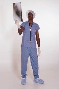
The most common and often the most expensive workers compensation claim is the back injury claim. Back injury claims often are associated with lifting heavier objects, twisting, bending, or falling. When the injured employee goes to the doctor, the doctor will normally treat the injury conservatively with rest and medication to see if the back injury is a sprain or strain of the musculoskeletal system.
If the injury does not respond to rest, the doctor will consider various types of diagnostic testing. There are several types of diagnostic testing the doctor can consider and request to determine the nature and extent of the back injury. The six most common diagnostic test are x-rays, MRIs, cat scans, EMGs, myelograms, and discograms.
1. X-rays
The most well known diagnostic test is the x-ray. It is often the first diagnostic test when the employee has fallen or suffered some other type of impact. The purpose of the x-ray is well known – to see if the injured employee has fractured any bones. The x-ray also allows the doctor to examine the vertebrae for other causes of back pain including osteoarthritis and for deformities like scoliosis.
The days of x-rays produced on film are no more. Today, x-ray images created by radiation are reproduced in a computer. There is very little risk to the employee in having an x-ray, unless the employee is pregnant, as radiation could harm a baby in utero. The results of x-rays are often available immediately, but there is usually a wait for the doctor to review the results.
Click Link to Access Free PDF Download
2. MRIs
Magnetic Resonance Imaging (MRI) is a way for the doctor to examine soft tissues in an employee’s body. The MRI machine uses radio waves, a large magnet and a computer to create images of soft tissue. In some cases, it is necessary for a dye to be inserted via a intravenous line (IV) into a vein to make it easier to detect inflammation or abnormality. Each MRI picture shows a view of the area being examined. Each picture is about a quarter inch deeper (or shallower) than the prior picture, allowing the doctor to get a detailed view of the area being examined. With a MRI of the spine, it shows areas where other structures may be impinging on nerve root areas. An MRI has no side effects, but occasionally there is a reaction to the dye.
The MRI machine is a circular tube with a table in the middle that the injured employee lays on — though response to claustrophobic patients has encouraged the creation of standing MRI machines. Typically, the MRI technician moves the table back and forth in and out of the tube while each MRI scan is taken. If dye is needed, it is injected about halfway through scanning. The employee will be told to hold their breath while each picture is taken. The MRI pictures are recorded on film which the MRI technician develops. It takes a doctor trained in reading the images to examine and interpret the images. Many physicians consider and MRI to be the best use as a pre-surgical tool.
3. CT ScansA Computed Tomography scan, referred to as a CT scan, or a Computerized Axial Tomography Scan, or CAT scan, is another way of taking pictures of the body using a specialized x-ray machine. The machine circles the employee’s body and scans the area from every angle. The machine measures how x-ray beams change as they pass through the body. A computer generates a series of black and white pictures each showing a slightly different cross section.
If the x-rays and the MRI have not identified the cause of the employee’s back pain, the doctor may request a CAT scan. The CAT scan is not often used for back injuries. When the treating doctor asks for a CAT scan instead of an MRI, the doctor is looking for some other reason the employee is experiencing back pain including problems with the kidneys and pancreas.
A CAT scan is done much the way a MRI is done. The employee lies on a table that passes in and out of the tube-shaped machine. The CAT scan is done with dye to outline the soft tissues and blood vessels. There is a small amount of risk from the dye. Some people have an allergic reaction to it. Also, since the CAT is a specialized x-ray machine, it should not be used on pregnant employees.
4.EMG
Electromyography (EMG) is used to test the employee’s nerve and muscle electrical activity. EMGs are usually done with a nerve conduction study (NCS). If the treating doctor suspects the back injury and resulting pain is to muscles or a pinched nerve, an EMG may be requested. In the EMG test, the employee has fine needles inserted into the muscles. Each needle is attached to a wire that sends signals to the EMG machine. The electrical patterns inside the muscles can be analyzed to determine which muscle is damaged. The EMG portion checks on muscle responses and the NCS checks nerve velocities. Together both are interpreted to help diagnose many problems from nerve
impingement to neuropthies and more.
EMG needles are too small to cause bleeding but most employees find the test uncomfortable. The electrical shocks that occur in the test are too mild to cause any permanent damage.
FREE DOWNLOAD: “5 Critical Metrics To Measure Workers’ Comp Success”
5. Myelogram
A myelogram is an x-ray combined with a dye that is injected directly into the spinal canal. The myelogram is used to identify the point(s) in the employee’s back where vertebrae are pinching the spinal cord. It is often used prior to surgery to confirm the MRI results. The myelogram is also used to diagnosis leg pain problems occurring in conjunction with back injury.
As with other dyes used in testing, some people have an allergic reaction. Also some people experience headaches from the dye, and pregnant women should not have the test done.
6. Discogram
Under monitored conditions, sterile water is injected into several adjacent disc spaces to attempt to reproduce symptoms (i.e., parasthesias, pain). This test is subjective but common preoperatively to help doctors make sure they are operating at the source if the pain — which is not always the “worst disc.”
Now when you hear the employee is having one of these tests done, you will have an idea what is happening to diagnose the employee’s back problem.
Author Rebecca Shafer, JD, President of Amaxx Risks Solutions, Inc. is a national expert in the field of workers compensation. She is a writer, speaker, and website publisher. Her expertise is working with employers to reduce workers compensation costs, and her clients include airlines, healthcare, printing, publishing, pharmaceuticals, retail, hospitality, and manufacturing. See www.LowerWC.com for more information. Contact: RShafer@ReduceYourWorkersComp.com.
Our WC Book: http://www.wcmanual.com
WORK COMP CALCULATOR: http://www.LowerWC.com/calculator.php
MODIFIED DUTY CALCULATOR: http://www.LowerWC.com/transitional-duty-cost-calculator.php
WC GROUP: http://www.linkedin.com/groups?homeNewMember=&gid=1922050/
SUBSCRIBE: Workers Comp Resource Center Newsletter
©2011 Amaxx Risk Solutions, Inc. All rights reserved under International Copyright Law. If you would like permission to reprint this material, contact Info@ReduceYourWorkersComp.com.
FREE DOWNLOAD: “5 Critical Metrics To Measure Workers’ Comp Success”


